Left Hip Muscles Anatomy : Muscle and tendon anatomy of the hip (adductors, gluteal muscles (or buttocks).. In clinical anatomy the thigh muscles are divided into three groups: Semi means half and tendinosus means tightly stretched band (otherwise known as a tendon). Anatomy of the muscular system. 3 months later i got acute excrutiating pain in inguinal area. There are a lot of muscles of the hip and thigh.
Rectus femoris forms the middle portion of the quadriceps. Each muscle below has the bones in bold for intermediate learners and the specific bony landmarks for advanced learners. The main muscles of the hip and pelvis consistsof the iliopsoas, pectinues, rectus femoris and sartorius at the front. The geometry of the hip allows wide range of motion in all planes. A bursa that sometimes causes problems in the hip is sandwiched between the bump on the outer hip (the greater trochanter) and the muscles and tendons that cross over the bump.
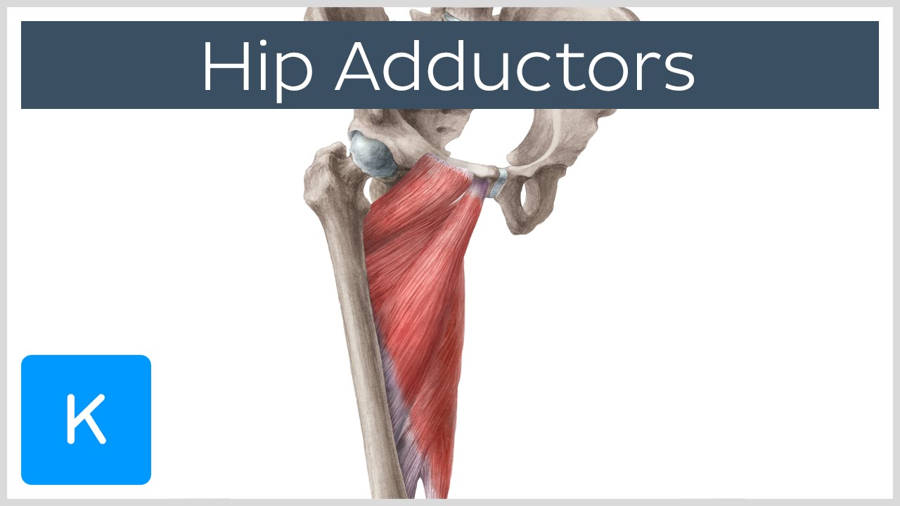
Anatomy of the Hip Adductor Muscles - Human Anatomy ... from i.ytimg.com Muscle and tendon anatomy of the hip (adductors, gluteal muscles (or buttocks). for detailed anatomy of pelvic bones, read anatomy of hip bone. Anatomy of the muscular system. Human anatomy hip muscles anatomy anatomy study. Left leg, lateral (left) and posterior (right) views. Each muscle below has the bones in bold for intermediate learners and the specific bony landmarks for advanced learners. Your email address will not be published. The geometry of the hip allows wide range of motion in all planes.
It is a flat, triangular muscle on the anterior wall of the pelvis.
The muscles of the pelvis, hip and buttock anatomical chart shows how each muscle in this area of the body works with the others, and the various minor systems within the major ones. Comprehensive information about hip joint anatomy including muscles, tendons, ligaments, bones, bursae, skeletal structure and joint capsules. Now that you watched the video, you. Related online courses on physioplus. This month's muscle of the month focusses on the hamstrings, a very powerful group of muscles that extend the hip and flex the knee. Anatomy of the muscular system. Most modern anatomists define 17 of these muscles, although some additional muscles may sometimes be considered. This anatomical atlas was especially designed for a specific public (radiologists, surgeons, rheumatologists and physicians specializing in musculoskeletal imaging). The cavity of the acetabulum the external obturator muscle is short external rotator muscle of hip joint. A bursa that sometimes causes problems in the hip is sandwiched between the bump on the outer hip (the greater trochanter) and the muscles and tendons that cross over the bump. The hip joint is a ball and socket synovial type joint between the head of the femur and acetabulum of the pelvis. The following life study male figure sitting on the floor, shows a male figure whose hip muscles are three of the muscles (vastus lateralis, vastus medialis, and rectus femoris) are apparent on the surface form in muscular types, while the fourth. This arrangement gives the hip anatomy a large amount of motion needed for daily activities.
Semimembranosus, semitendinosus and biceps femoris (the hamstrings). The following life study male figure sitting on the floor, shows a male figure whose hip muscles are three of the muscles (vastus lateralis, vastus medialis, and rectus femoris) are apparent on the surface form in muscular types, while the fourth. 3 months later i got acute excrutiating pain in inguinal area. The hip muscles encompass many muscles of the hip and thigh whose main function is to act on the thigh at the hip joint and stabilize the pelvis. Pelvis and acetabulum, with muscle attachment sites.
Female Torso Musculature Labelled Back Muscles Anatomy ... from i.pinimg.com Muscles that act on the lower limb cause movement at the hip, knee and foot joints. Learn their anatomy efficiently and easily using kenhub's muscle anatomy and reference charts! 3 months later i got acute excrutiating pain in inguinal area. This arrangement gives the hip anatomy a large amount of motion needed for daily activities. If left unstretched, shortened hip flexors affect the position of the pelvis, which in turn affects the position and movement of the lower back. The cavity of the acetabulum the external obturator muscle is short external rotator muscle of hip joint. In utero fetal hips lie typically in flexion, abduction and external rotation, with the left hip usually muscular anatomy. If you know all the hip flexor names and bones they attach to, that's an awesome accomplishment!
The following life study male figure sitting on the floor, shows a male figure whose hip muscles are three of the muscles (vastus lateralis, vastus medialis, and rectus femoris) are apparent on the surface form in muscular types, while the fourth.
Left leg, lateral (left) and posterior (right) views. for detailed anatomy of pelvic bones, read anatomy of hip bone. Muscles that act on the lower limb cause movement at the hip, knee and foot joints. 3 months later i got acute excrutiating pain in inguinal area. It is a flat, triangular muscle on the anterior wall of the pelvis. The geometry of the hip allows wide range of motion in all planes. The hip's unique anatomy enables it to be both extremely strong and amazingly flexible, so it can bear weight and allow for a wide range of movement. The hip joint is the articulation of the pelvis with the femur, which connects the axial skeleton with the lower extremity. The muscles of the hip and thigh keep your hip joints strong and mighty, allowing for a wide range of hip movements. The hip muscles encompass many muscles of the hip and thigh whose main function is to act on the thigh at the hip joint and stabilize the pelvis. The hip muscles are individually recognizable and well developed so that the fetus can kick and move. Anatomy, bony pelvis and lower limb, psoas major. Knee assessment and hip mechanics online course:
Now that you watched the video, you. Semimembranosus, semitendinosus and biceps femoris (the hamstrings). Muscles that act on the lower limb cause movement at the hip, knee and foot joints. Your email address will not be published. Learn their anatomy efficiently and easily using kenhub's muscle anatomy and reference charts!
Muscles of the hips and thighs | Human Anatomy and ... from s3-us-west-2.amazonaws.com Knee assessment and hip mechanics online course: Two individual muscles called the psoas major and the iliacus form the iliopsoas muscle. Learning the anatomy of your hip will better enable you to pinpoint your pain and work with your doctor to keep it from limiting your life. Semimembranosus, semitendinosus and biceps femoris (the hamstrings). This muscle is about half tendinous. Learn about anatomy hip muscle with free interactive flashcards. Human anatomy hip muscles anatomy anatomy study. The geometry of the hip allows wide range of motion in all planes.
Knee assessment and hip mechanics online course:
1 hip anatomy, function and common problems. Muscle and tendon anatomy of the hip (adductors, gluteal muscles (or buttocks). Comprehensive information about hip joint anatomy including muscles, tendons, ligaments, bones, bursae, skeletal structure and joint capsules. If you know all the hip flexor names and bones they attach to, that's an awesome accomplishment! Anatomical terms allow us to describe the body and body motions more precisely. Knee assessment and hip mechanics learn how hip. Advanced hip flexor muscle anatomy. Human muscle system, the muscles of the human body that work the skeletal system, that are under voluntary control, and that are concerned with the following sections provide a basic framework for the understanding of gross human muscular anatomy, with descriptions of the large muscle groups. In human anatomy, the muscles of the hip joint are those muscles that cause movement in the hip. In clinical anatomy the thigh muscles are divided into three groups: The fibers of this muscle attach to the lower eight ribs and spiral downward and medially to attach to the hip bone. Microscopic anatomy of skeletal muscle. Related online courses on physioplus.


0 Comments